Through the years, ocular pathology has added value to the practice of ophthalmology by structurally defining and understanding disease processes and helping make ophthalmology the medical and surgical specialty it is today.[1] A/Prof Sonja Klebe is a surgical pathologist at Flinders Medical Centre SA with a special interest in ocular pathology and explains more about this discipline.
“Ocular pathology deals with the nature and causes of diseases of the eye and its surrounding structures, their effect on the ocular tissues and ocular functions, and subsequent management options. Traditionally, ocular pathology was performed by ophthalmologists, however now it has grown into a subspecialty of its own, established under both ophthalmology and surgical pathology.
“It is essential that the final histopathology report is accurate and contains as much relevant information as possible for safe patient management, including diagnosis and subsequent treatment options. Extensive knowledge of the underlying eye conditions is therefore absolutely critical, plus a strong working relationship with the ophthalmologist,” said A/Prof Klebe.
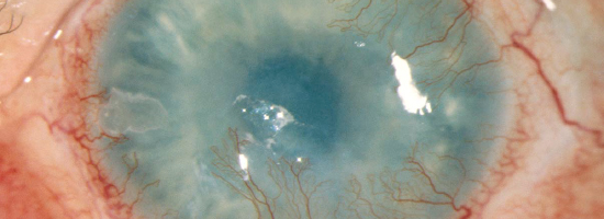
Ocular pathologists make clinical diagnoses through the evaluation of tissue, by gross examination (describing and measuring the tissue), inking (if required), sectioning the tissue to be processed for diagnosis, and routine histopathology and special stains.
“Ocular pathology can be quite challenging. Not only are there a wide range of tumours which require molecular characterisation to determine therapy and prognosis, there is also a whole spectrum of skin diseases relating to the lids of the eye. Because of this, the different disciplines are required to work together, including molecular pathology, genetics, microbiology and anatomical pathology. Assessment of corneal dystrophies is also one of the areas where electron microscopy (EM) still forms the standard of assessment.
“Very specialised surgical techniques are required to take specimens from a patient. For example, there may be eight or nine different specimens just relating to the cornea alone, which all have to be handled in a completely different way. The biopsies are also extremely small, which is particularly challenging when processing as they need to be divided into several parts. As they are often transparent and put into a clear media, the first challenge is to find the specimen!”
“There needs to be a lot of trust between the pathologist and the clinician to ensure biopsy the right area. It is very easy to get that wrong. If you get that wrong it can mean that a young person loses their eyesight or alternatively, a young person goes on to develop a tumour which ultimately kills them. Ocular pathology is one of the areas where the different subspecialties of pathology work effectively together for the benefit of the patient,” said A/Prof Klebe.